
α-Naphthyl Acetate Esterase (α-NAE) Stain
Intended Use:
This kit is for staining bone marrow cell and bloodcell smear.
Principle:
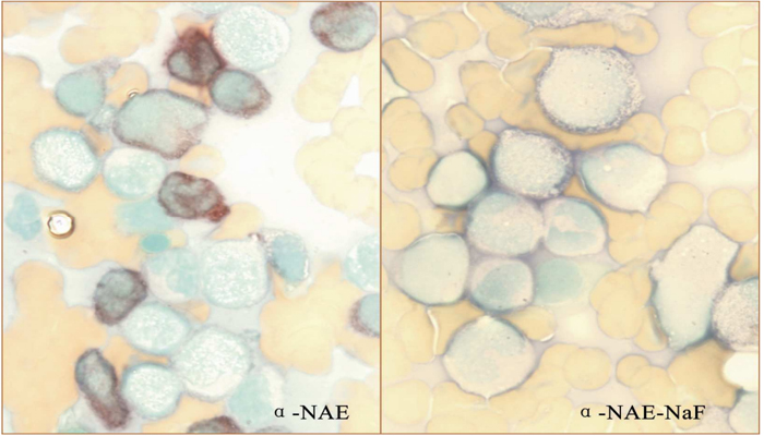
Hydrochloric acid parafuchsin reacts with Sodium Nitrite to form diazonium salt (six-azo-pararosaniline). α-Naphthyl Acetate is decomposed by esterase and produces α-naphthol, which shall combine with diazonium salt to form red brown precipitate in cytoplasm. This stain has no specificity against esterase; therefore, it is called "Non-specific Esterase Stain".
Specifications:
|
Contents |
5Tests/Kit |
20Tests/Kit |
100Tests/Kit |
Components |
|
Fixative(solution A) |
1vial×2.5ml |
1vial×10ml |
1vial×50ml |
Sodium Citrate |
|
Pararosaniline |
1vial×0.3ml |
1vial×1.2ml |
1vial×5.5ml |
Parafuchsin |
|
Sodium Nitrite |
1vial×0.3ml |
1vial×1.2ml |
1vial×5.5ml |
Sodium Nitrite |
|
PhosphateBuffer |
1vial×8ml |
2vials×18ml |
2vials×90ml |
Phosphate |
|
α-Naphthyl Acetate |
1vial×0.3ml |
1vial×1.2ml |
1vial×5.5ml |
α-Naphthyl |
|
Methyl Green |
1vial×5ml |
1vial×20ml |
1vial×100ml |
Methyl Green |
Methods:
Working solution Preparation (for 1 test only):
Apparatus required: Disposable tube, micropipette, disposable tips, dropper;
Instruction: Add 50µl solution B and solution C in a test tube ; mix well and wait for 1 minute. Then, add 1.5ml solution D and 50µl solution E; mix well and wait for 2 minutes. (Add 1 drop of solution NaF for NaF inhibition test.)
| Solution B | Solution C | Solution D | Solution E | Solution NaF | |
| Tube Α | 50µl | 50µl | 1.5ml | 50µl | -- |
|
Tube B (inhibition test) |
50µl | 50µl | 1.5ml | 50µl | 1 drop |
Expected Results:
Red or brown granules found in cytoplasm are positive.
ó┘Spot pattern: It is mainly seen in mature T Lymphocytes. 1~4 brown or red-brown circular, mass, and big spot granules with clear contour in cytoplasm are found in positive reaction.
ó┌Diffuse pattern: Red-brown and dusty granules diffusely spread are found in positive reaction. Granules
may appear at a certain part of cell with obscure outline in cytoplasm.
ó█Monocyte Pattern: Evenly stained red-brown granules diffusely spread in cytoplasm are found in
positive reaction.